

In addition to the fact that it is a moment that fills you with hope, because you will be able to see your baby and listen to his heart, the ultrasound at week 12 is also one of the most important during pregnancy from a medical point of view.
If the time has come to have it done and you have doubts, don’t worry, in this article we will explain everything, from why it is so important and what you can know with it, to why the doctor has also scheduled a blood test for you And how do you have to be prepared?
WHAT IS THE 12 WEEK ULTRASOUND?
The 12-week ultrasound is one of the most important in the assessment and monitoring of pregnancy (along with the 20 -week and 36 -week ultrasounds ). It is carried out between weeks 11 and 13 , a stage in which the fetus is in full formation of all its systems and organs and is already large enough to be able to appreciate its development.
This ultrasound provides very relevant data on the development of the baby that can be key to the detection of possible chromosomal and morphological abnormalities , among other things.
WHAT IS THE GOAL OF THE 12 WEEK ULTRASOUND?
As we have mentioned, the main function of this test is to observe how the baby is doing and detect possible developmental problems in time.
Through this ultrasound, the following information can be obtained:
- Check gestational age. Although pregnancy is counted from the date of the last period, not all women ovulate at the same time of the cycle, so this test is used to estimate the real time of gestation calculated according to the skull-rump or skull-tail length of the fetus.
- Confirm embryonic vitality. It checks if there is a heartbeat.
- Know the number of embryos. In this ultrasound it is possible to know if it is a single embryo or a multiple pregnancy . And, in the latter case, how many embryos are there and if they share the placenta or the sac.
- Check the risk of chromosomal abnormalities . Among other things, the sonographer will observe and measure the nuchal fold, to later cross this data with others in the triple screening and thus calculate the risk of chromosomal abnormalities.
- Detect morphological abnormalities . Although it seems early, the baby is already forming and, therefore, there are already some abnormalities that may be detectable: ventriculomegaly, holoprosencephaly, anencephaly, structural heart disease, omphalocele, renal agenesis, megabladder, facial changes and changes in the size of the limbs.
- Detect the risk of preeclampsia. Preeclampsia is a pregnancy disease that is caused by a problem (not yet identified) during the formation of the placenta and manifests itself in high blood pressure and alterations in various organs of the body, while affecting the supply of oxygen and nutrients to the placenta and also the growing baby.
IS IT DONE ABDOMINALLY OR VAGINALLY?
Being an early stage in pregnancy, this ultrasound can be carried out both vaginally and abdominally.
The difference between the two methods is that:
- With the probe in the abdomen, larger planes can be obtained, but with poor resolution.
- While, when doing it vaginally, the planes are more limited (they are conditioned to the path of the probe), however, the images are much clearer.
The decision of which system to use depends on the sonographer and, sometimes, if he considers it appropriate, he can even combine both.
WHAT IS TRIPLE SCREENING?
The triple screening, also called chromosomal abnormality screening, is a study that is carried out together with the ultrasound at week 12. To do this, coinciding with the ultrasound, the doctor will request a blood test.
This screening consists of an estimation of the probabilities that the fetus is affected by Down Syndrome (trisomy 21), Edwards Syndrome (trisomy 18) or Patau Syndrome (trisomy 13), whose calculation is made taking into account three indicators:
- The age of the mother , since the older she is, the more risk there is.
- Blood levels of the hormone free beta-hCG and the protein PAPP-A , manufactured by the placenta.
- nuchal translucency . During the ultrasound, the specialist measures the thickening of the nuchal fold, located at the back of the fetus’s head.
The triple screening, however, is only a mathematical calculation that measures a risk index, it does not provide conclusive results or establish a diagnosis .
In any case, if this index tells us that the risk is high (if it is greater than or equal to 1/250 for trisomy 21, 13 or 18), other more precise tests would be carried out that could rule out or confirm the risk. risk such as a fetal DNA test in maternal blood or invasive tests such as amniocentesis or chorionic biopsy.
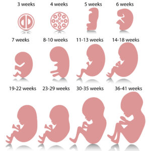
WHAT DOES A 12-WEEK EMBRYO LOOK LIKE?
At 12 weeks, your baby’s appearance begins to resemble that of a newborn. His vital organs have already formed and you will be able to distinguish his head, which is beginning to round, his thorax, his abdomen and his extremities (feet and hands are already developing).
At this stage it measures between 60 and 70 millimeters and weighs approximately 9 to 14 grams (it would fit in the palm of your hand). It is surrounded by a large amount of amniotic fluid and it moves a lot, you may see it kicking, shaking its arms or even its little head.
WILL YOU TELL ME IF THE BABY IS A BOY OR A GIRL?
At 12 weeks the baby’s genitals have already finished forming, so there are chances that in this ultrasound you can already know their sex. However, on some occasions it may not be easy to see it or what we think we see may not be entirely reliable.
DO I NEED ANY KIND OF PREPARATION FOR THE FIRST TRIMESTER ULTRASOUND?
The ultrasound at week 12 does not require any special preparation, as long as the doctor does not tell you otherwise.
Keep in mind that although the tests to measure B-HCG and PAPP-A do not require fasting, it may be the case that other measurements that do require fasting are included. To be sure, we advise you to consult it.
However, if nothing else is indicated, the only recommendation for the ultrasound is that you do not apply creams, oils or lotions to your abdomen for 48 hours before the test, because they could interfere with the test.
CONCLUSIONS
The 12 week ultrasound is one of the most important pregnancy tests. This test allows the status and development of the fetus to be observed and, combined with the triple screening, allows the detection of possible chromosomal abnormalities.
In addition, it is the first in which you will have the opportunity to see your baby with an appearance more similar to a newborn (although much smaller), listen to his heartbeat and, if possible and if you want, know his sex.
Average Rating